MFM (magnetic force microscopy) is a type of SPM (scanning probe microscopy).
If you would like to know more about SPM, please read the following article, which also explains it.

This article provides a simple explanation of how MFMs observe samples.
We at Suga, who work with atoms on a daily basis, are uniquely positioned to share this information. We hope you find it useful.
Let’s take a quick look.
Contents
1 MFM (magnetic force microscopy) can image the distribution of magnetic force gradients.

MFM is a type of microscope capable of imaging the distribution of magnetic force gradients.
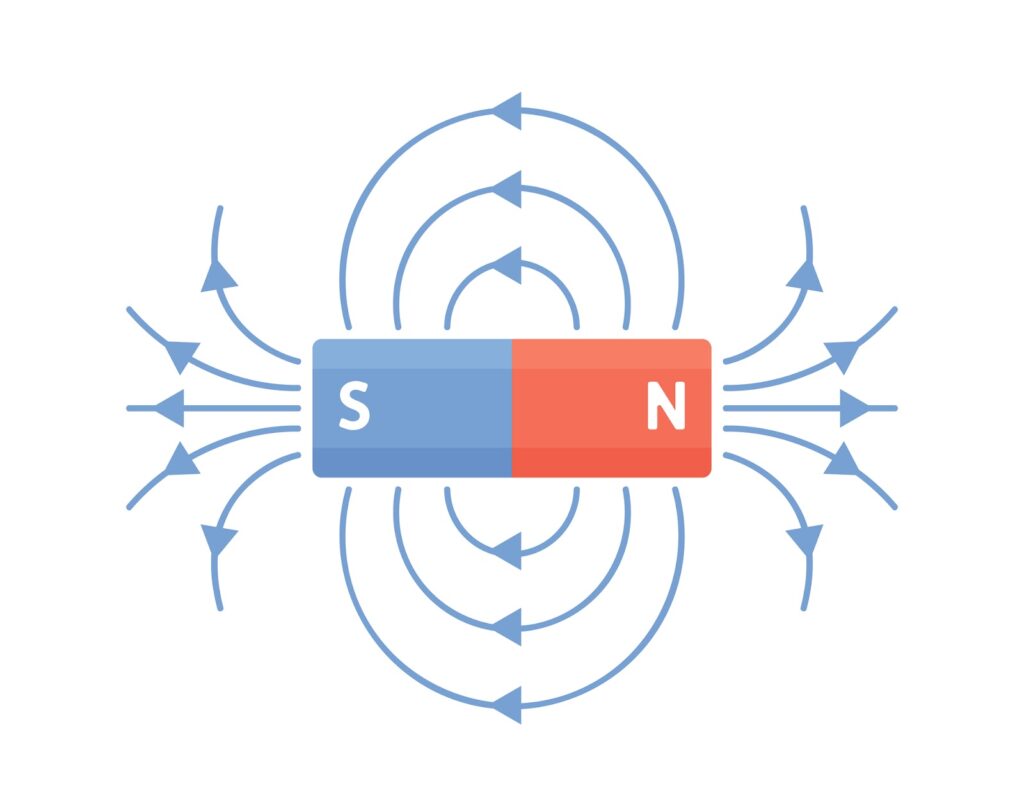
In simple terms, it allows you to visualize objects with magnetic force. (it can make images of gravitational and repulsive forces.)
Magnetic force microscopy (MFM) is a type of non-contact atomic force microscopy (NC-AFM) that uses the cantilever employed in AFM, with a magnetic film coating applied to the probe.
As a development of AFM, it has the characteristics of being able to make measurements in air, non-contact and non-destructive.
Sputter deposition is also used to coat this magnetic film.
For more information on sputter deposition, see the following article.

So how does MFM work and how does it observe a sample? See the followings.
Structure of MFM (magnetic force microscopy)
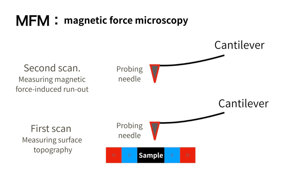
The MFM detects two main measurements.
The two targets are the cantilever’s motion and the magnetic interaction between the probe and the sample.
First, the surface profile is measured and its movement is recorded.
While tracing the movement of the cantilever, the next step is to measure the amplitude of the run-out caused by the magnetic force, and the MFM outputs an image with a mechanism that ascertains the distribution of magnetic forces by producing a phase change.
The measurement of cantilever movement is similar to the AFM mechanism.

Imaging the forces of attraction and repulsion acting between the MFM probe and the magnetic sample produces an image, called an MFM image.
The next section presents a sample of real images.
Real image samples from MFM (magnetic force microscopy)
The real image of the MFM can be seen before and after data erasure on the hard disk.
Black is the N-pole and white is the S-pole, and when four are solidified, they form a 4-bit magnetic signal.
Before erasure, the data is present and stored as a bit signal; after erasure, it is gone.
For a sample of the actual image, please see the following page.
MFM microscopy to confirm erasure.
Application of MFM (magnetic force microscopy)
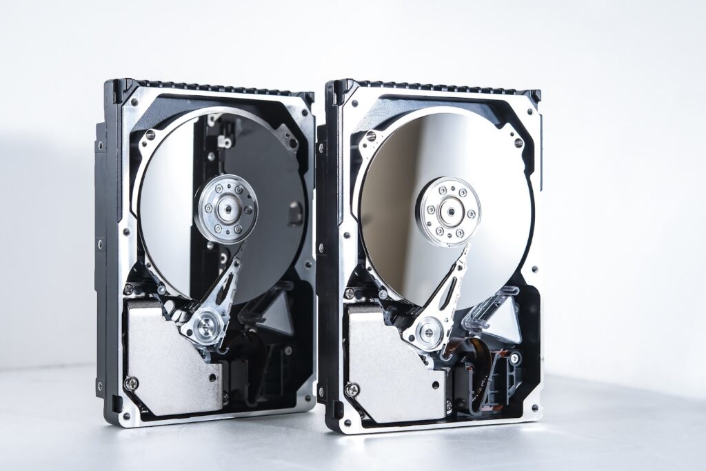
The most famous application of MFM technology is the ‘hard disk’.
As mentioned earlier, hard drives that used N and S poles and recorded as bits were widely used, particularly in the PC sector.
Recently, the widespread use of SSDs has ceded their place of activity.
The MFM itself is also used for observations in the biotechnological field, such as magnetic bacteria.
History of MFM (magnetic force microscopy)

Research into MFM began in the 1980s.
When it was first developed, it was difficult to create magnetic chips and was beyond the scope of research.
However, with the birth of photolithography technology and next deposition coating technology, it became practical in the early 1990s.
Even today, this microscope continues to play an active role as one of the SPMs for the elucidation of quantum properties.
Summary
We talked about MFM (magnetic force microscopy) in this issue.
Finally, let’s look back at the key points.
・MFM (magnetic force microscopy) can image the distribution of magnetic force gradients.
・Sputter deposition of a magnetic film on the probe of an AFM (Atomic Force Microscope) to enable it to sense magnetic forces.
・The mechanism is that the shape is measured in the first session and the movement caused by the magnetic force is measured in the second session, and the distribution of the magnetic force is imaged by producing a phase change.
・It was often used in hard disks and other recording media.
・Now, they are active in biotechnological observations and in the elucidation of quantum properties.
We mentioned at the beginning that MFM is a type of SPM (scanning probe microscope).
For more information on SPM, see also the following article.
References site
磁気力顕微鏡(MFM:Magnetic Force Microscope) : 日立ハイテク
7-1 磁気力顕微鏡 (MFM) – 原子間力顕微鏡 (AFM) の仕組み | How AFM Works
磁気力顕微鏡の原理と応用
第107回「ナノテクを結集した磁気力顕微鏡」の巻
https://www.jstage.jst.go.jp/article/jsssj1980/17/1/17_1_8/_pdf
産総研:世界最高分解能の磁気力プローブ顕微鏡を開発