SNOM (scanning near-field optical microscope) is a type of SPM (scanning probe microscope).
If you are interested in SPM, please read the following article.
This article will explain some examples of how SECMs work and the situations in which they are used.
We are pleased to share insights that only those of us working with atoms on a daily basis can provide. We hope you find this information useful.
Let’s look at it.
Contents
SNOM is a scanning microscope that uses a special type of light called near-field light.

Near-field light has the property that it does not travel through space and disappears halfway through, making it invisible.
Observation is therefore carried out by scanning the sample using a thin probing needle.
One advantage is that measurements can be made in air and non-destructively because it is not just a scanning probe microscope but also an optical microscope.
However, there are disadvantages, such as inferior resolution compared to scanning tunneling microscopy.
SNOMs are used to detect gene positions on DNA and to measure chromosome surface structure.
Structure of the SNOM (scanning near-field optical microscope).
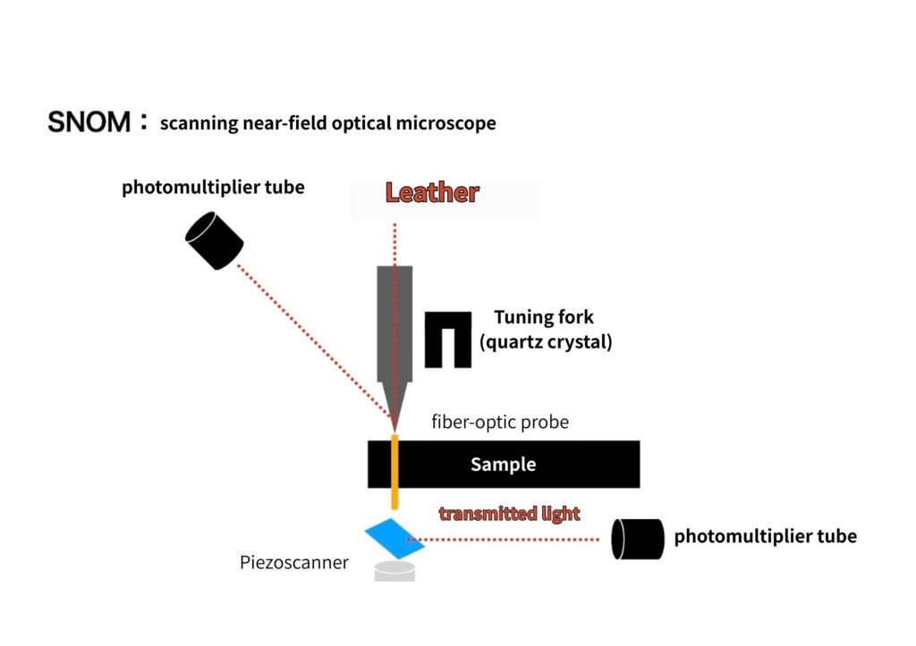
SNOM (scanning near-field optical microscope) images structures by vibrating a tuning fork (quartz crystal) fitted with a fiber-optic probe with a small hole at its tip and detecting amplitude changes when the probe tip contacts the material.
Imagine a scanning probe microscope that uses light instead of atomic forces or tunneling currents for detection.
Reference: SNOM – Scanning near-field optical microscope.
Real image samples from SNOM (scanning near-field optical microscope).
The real image of “alpha300S” by “OXFORD INSTRUMENTS” showed the SNOM image of the metaphase of the human chromosomes.
Chromosomes are on the nanoscale, but this microscope is so good that the shapes are clearly visible when zoomed in.
SNOM image of a metaphase of a human chromosome.
The shapes are clearly visible, even though on the nanoscale.
SNOM images of surface plasmon reactions excited by nanoscale metal grids can also be seen, and other real image samples unique to SNOMs working in the nano field can be seen
Plasmon is explained in the following article for those interested.

History of SNOM (scanning near-field optical microscope)

The SNOM is not developed by a single researcher, but the result of multiple studies.
The first imaging report of SNOMs, then called NFOs, was made in 1985.
Separately from that report, the SNOM was developed by Fischer in Germany.
In addition, Cornell and his group at Bell Labs in the USA have developed their own SNOM.
With the above in mind, a highly practical SNOM was realized by Betzig et al. in 1991 and is used in research today.
Summary
We talked about SNOM (scanning near-field optical microscopy).
Let’s look back at the key points.
・SNOM (scanning near-field optical microscope) is a microscope that uses near-field light.
・SNOM uses a fiber-optic probe to control the distance between the sample and the probe by vibrating the tuning fork and imaging.
・SNOM was devised by a number of researchers.
We mentioned at the beginning that SNOMs are a type of SPM (scanning probe microscope).
For more information on SPM, see also the following article.
References site
走査型近接場光学顕微鏡(SNOM:Scanning Near-field Optical Microscope) : 日立ハイテク
近接場光学顕微鏡(SNOM) – WITec Raman Imaging
SNOM – 走査型近接場光学顕微鏡
9-1 走査型近接場光学顕微鏡 (SNOM) – 原子間力顕微鏡 (AFM) の仕組み | How AFM Works
走査型近接場顕微鏡の表面化学への応用
走査型近接場光学顕微鏡 | オプティペディア – Produced by 光響
近接場光学顕微鏡/走査型 トンネル 顕微鏡複合装置の開発
近接場
SNOM/AFMの 生物試料への応用
【走査型近接場光顕微鏡】 | デジタルマイクロスコープなら朝日光学
alpha300 S – SNOM 走査型近接場光学顕微鏡 – WITec Raman Imaging
近接場磁気光学顕微鏡の現状と課題
【走査型近接場光顕微鏡から生まれた顕微鏡】